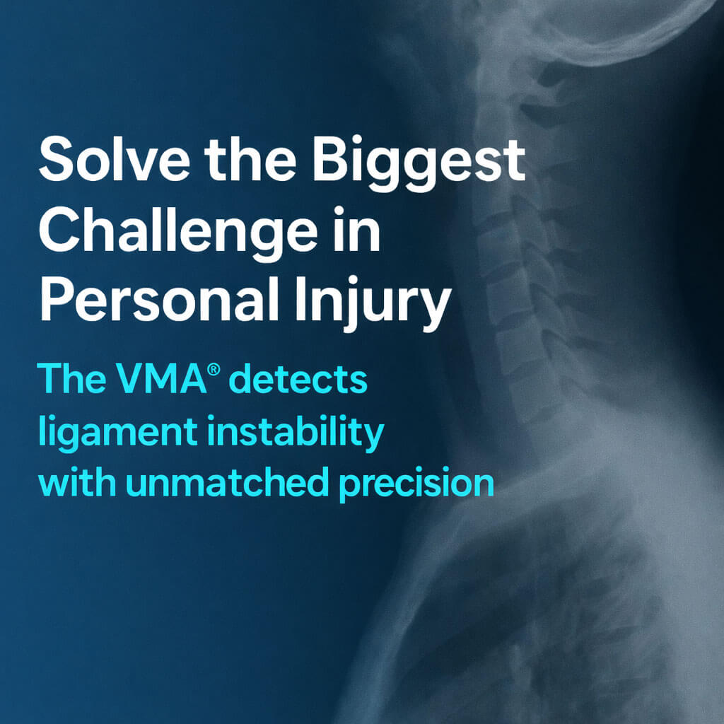
The Technology Behind the VMA®
Motion-Based Spinal Imaging Built for Objective Diagnosis
See Motion Measured
Real time motion
Visualizes spinal movement in motion.
90%
Consistent motion capture across standardized studies.*
0.5mm
Defined spinal translation measurement precision.*
FDA
Cleared motion-based imaging system
Where Motion-Based Diagnosis Matters Most
-
1
Moves beyond sprain and strain classifications
-
2
Documents instability not visible on MRI or X-ray
-
3
Provides objective, defensible diagnostic evidence
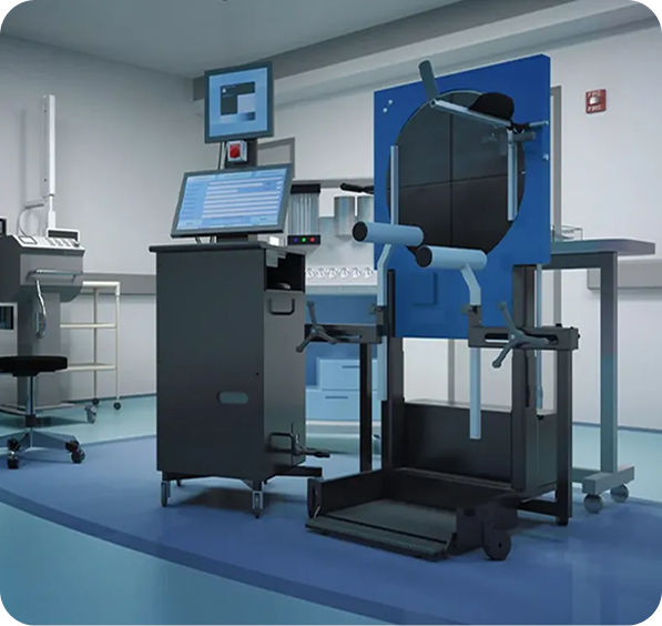
How the VMA Measures Spinal Motion
Weight-Bearing
Real-Time
Quantified
Repeatable
The Difference Is Motion
Why Motion-Based Imaging Changes Spinal Diagnosis
01
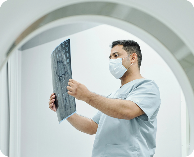
Static Images Miss Functional Injury
Ligament instability may remain undetected when the spine is evaluated only at rest.
02
The Spine Is a Dynamic System
True spinal behavior is expressed through movement, not static positioning.
03
Weight-Bearing Reveals Instability
Evaluating the spine under load reflects how patients function in daily life.
04
Motion Must Be Quantified
Objective diagnosis requires precise measurement, not visual interpretation alone.
05
Measurement Creates Diagnostic Confidence
Quantified motion data reduces uncertainty and strengthens clinical decision-making.
06
Objective Findings Support Better Care
When instability is clearly identified, diagnosis, documentation, and treatment align more effectively
System Expansion
- Integrate with clinical operations
- Streamline data handling
- Support consistent evaluations
Supporting Technologies
Tools That Extend
Motion-Based Diagnosis
- Enhance motion visualization
- Support quantitative assessment
- Aid diagnostic interpretation
A Diagnostic Technology Built for How the Spine Actually Works
- Captures spinal motion under real-world conditions
- Quantifies instability with precise measurement
- Produces standardized, defensible diagnostic reports
- Supports confident clinical and diagnostic decisions
Next Step
Book Your Strategy Session
Discuss whether motion-based spinal imaging is clinically and operationally appropriate for your practice.